Anatomy Of Ribs / The Intercostal Muscles of the Ribcage / Other classifications subdivide ribs into typical and atypical.. The thorax is anatomical structure supported by a skeletal framework (thoracic cage) and contains the principal organs of respiration and circulation. Any one of the paired bones, 12 on either side, extending from the thoracic vertebrae toward the median line on the ventral aspect of the trunk. A common complication of a rib fracture is they also have a role in ventilation; Bone structure of rib (c0035561). 12 photos of the anatomy of ribs and its related area.
Choose from 500 different sets of flashcards about anatomy ribs on quizlet. The first seven are connected behind with the vertebral column. Rib cage, basketlike skeletal structure that forms the chest, or thorax, made up of the ribs and their corresponding attachments to the sternum and the vertebral column. There are twelve (12) pairs of ribs and all articulate posteriorly with the thoracic vertebrae. They are ribbon like, elastic bony arches and flat in shape.
ribs anatomy from m.media-amazon.com There are twelve (12) pairs of ribs and all articulate posteriorly with the thoracic vertebrae. It protects the intercostal space containing the , , and. Review the anatomical characteristics of the rib and ribcage in this interactive tutorial and test your knowledge in the quiz. The final two pairs of ribs are floating ribs and the cartilage of these ribs tends to end ibrahim, af and darwish: The rib cage surrounds the lungs and the heart, serving as an important means of bony protection for these vital organs. Ribs 2 through 7 have a more traditional appearance and become longer and less curved as they progress downwards. There are two types of ribs, namely typical and atypical. The ribs stretches posteriorly from thoracic vertebrae to the anterior lateral edges of the sternum.
It protects the intercostal space containing the , , and.
The rib cage surrounds the lungs and the heart, serving as an important means of bony protection for these vital organs. The rib cage has a shape that resembles a cone briefly grows inferiorly as wide and form a hedge whose main functions are: The costotransverse ligaments in human: Protect the vital organs of the chest cavity as the heart, lungs and major blood vessels. Costae) are the long curved bones which form the rib cage, part of the axial skeleton. Curve by radius, curve by the edge. These three types can then be classified as either typical or atypical. One facet articulates with the numerically corresponding vertebrae. The final two pairs of ribs are floating ribs and the cartilage of these ribs tends to end ibrahim, af and darwish: There are twelve (12) pairs of ribs and all articulate posteriorly with the thoracic vertebrae. Vestibular anatomy and neurophysiology online course: Bone structure of rib (c0035561). Rib fractures most commonly occur in the middle ribs, as a consequence of crushing injuries or direct trauma.
In most tetrapods, ribs surround the chest, enabling the lungs to expand and thus facilitate breathing by expanding the chest cavity. There are two types of ribs, namely typical and atypical. Rib anatomy, thoracic rib, rib bone. The vertebral attachment of rib 1 can be found just below the neck and found above the level of the clavicle. The ribs are elastic arches of bone, which form a large part of the thoracic skeleton.
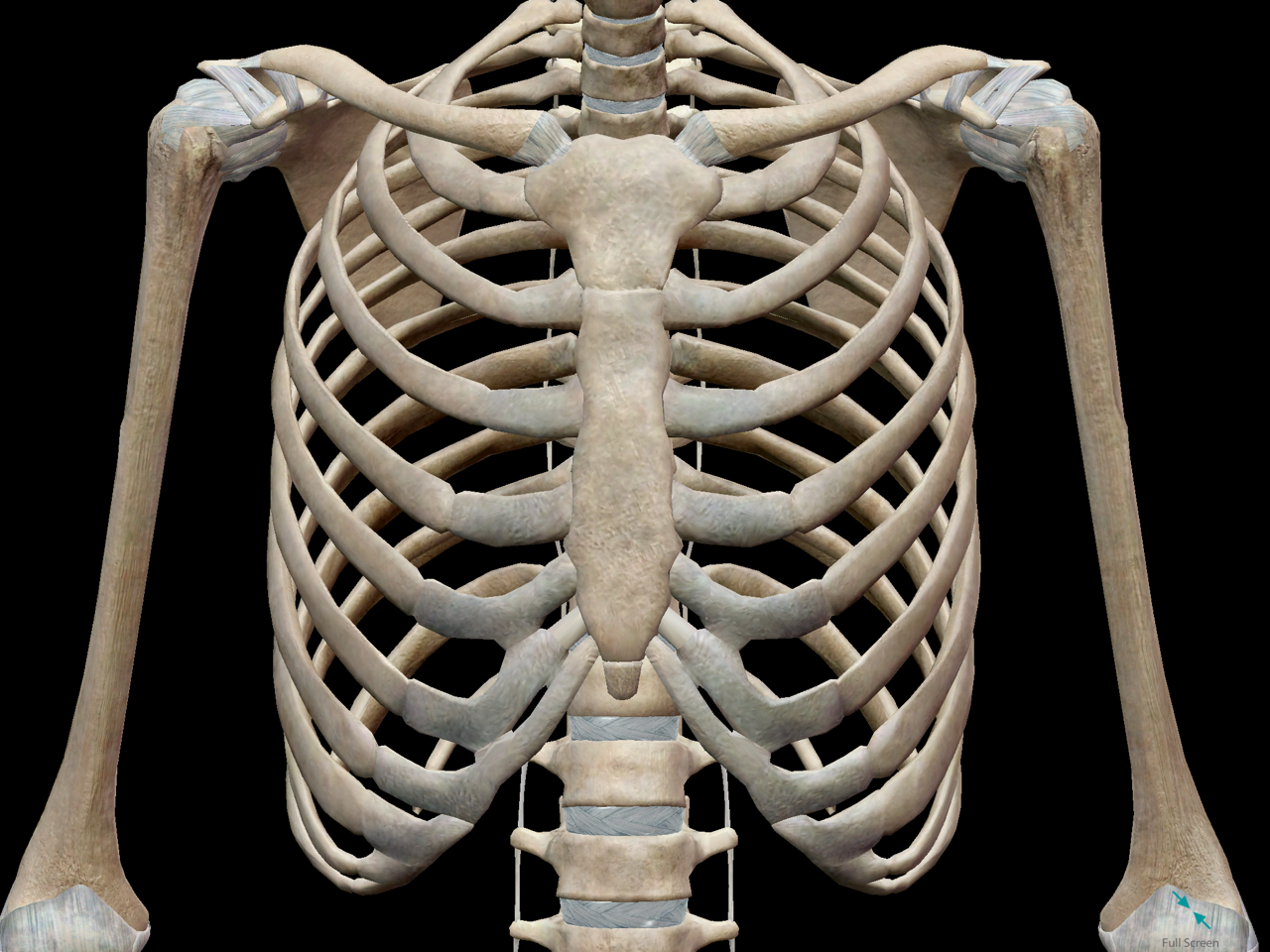
3D Skeletal System: Bones of the Thoracic Cage from www.visiblebody.com Individual ribs have a bony dorsal part, a body of rib, and ventral costal cartilage. These three types can then be classified as either typical or atypical. It protects the intercostal space containing the , , and. Bone structure of rib (c0035561). Introduction to the structure of the ribcage and ribs: The ribs stretches posteriorly from thoracic vertebrae to the anterior lateral edges of the sternum. They serve to protect the lungs, heart, and other internal organs of the. Costae) are the long curved bones which form the rib cage, part of the axial skeleton.
Vestibular anatomy and neurophysiology online course:
Ribs eight to ten are the false ribs and are connected to the sternum indirectly via the cartilage of the rib above them. Basic anatomy of ribs the rib cage, or thoracic cage, is a bony/cartilaginous structure surrounding the thoracic cavity and supporting the pectoral girdle. Major landmarks of a typical rib are the following: Introduction to the structure of the ribcage and ribs: They are ribbon like, elastic bony arches and flat in shape. The sternum and rib cartilage to ensure the ribs to the sternum above. Learn about anatomy ribs with free interactive flashcards. Includes images, video, and free quiz. Vestibular anatomy and neurophysiology online course: Typical ribs have a normalized general structure, while atypical ribs ribs three through nine are considered the typical ribs and are alike in structure and function. Learn about rib cage anatomy from spinal expert sarah key. The vertebral attachment of rib 1 can be found just below the neck and found above the level of the clavicle. The ribs help protect vital organs in the thorax such as the heart.
Bone structure of rib (c0035561). In most tetrapods, ribs surround the chest, enabling the lungs to expand and thus facilitate breathing by expanding the chest cavity. They are ribbon like, elastic bony arches and flat in shape. Costae are arranged in pairs and articulate with two successive vertebrae. Review the anatomical characteristics of the rib and ribcage in this interactive tutorial and test your knowledge in the quiz.
ribs anatomy from m.media-amazon.com A typical human rib cage consists of 24 ribs, the sternum (with xiphoid process), costal cartilages. The ribs stretches posteriorly from thoracic vertebrae to the anterior lateral edges of the sternum. The costotransverse ligaments in human: There are twelve pairs of ribs, all of which articulate ribs 3 to 9 are considered typical ribs. In vertebrate anatomy, ribs (latin: Major landmarks of a typical rib are the following: Protect the vital organs of the chest cavity as the heart, lungs and major blood vessels. Coastal cartilages are joined to the.
Rib fractures most commonly occur in the middle ribs, as a consequence of crushing injuries or direct trauma.
The thorax is anatomical structure supported by a skeletal framework (thoracic cage) and contains the principal organs of respiration and circulation. The ribs, along with the thoracic vertebrae, sternum, and costal cartilages, make up the thoracic cage, also. Coastal cartilages are joined to the. They increase in length, curvature and amount of cartilage craniocaudally. Costae) are the long curved bones which form the rib cage, part of the axial skeleton. A common complication of a rib fracture is they also have a role in ventilation; But this number may be increased by the development of a cervical or lumbar rib, or may be diminished to eleven. There are two types of ribs, namely typical and atypical. The ribs are elastic arches of bone, which form a large part of the thoracic skeleton. The final two pairs of ribs are floating ribs and the cartilage of these ribs tends to end ibrahim, af and darwish: The first seven are connected behind with the vertebral column. Individual ribs have a bony dorsal part, a body of rib, and ventral costal cartilage. Costae) are long, flat, curved bones that form the rib cage.


0 Komentar